| Brand/Manufacturer | GE Healthcare |
|---|---|
| SKU | GE-VIVID-EXPLORER |
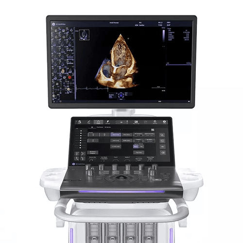
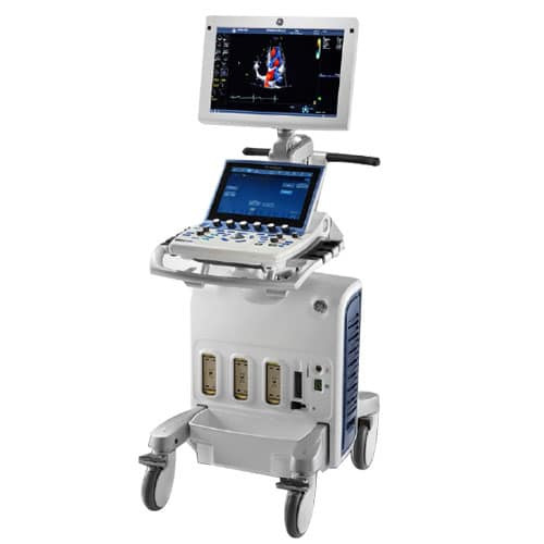
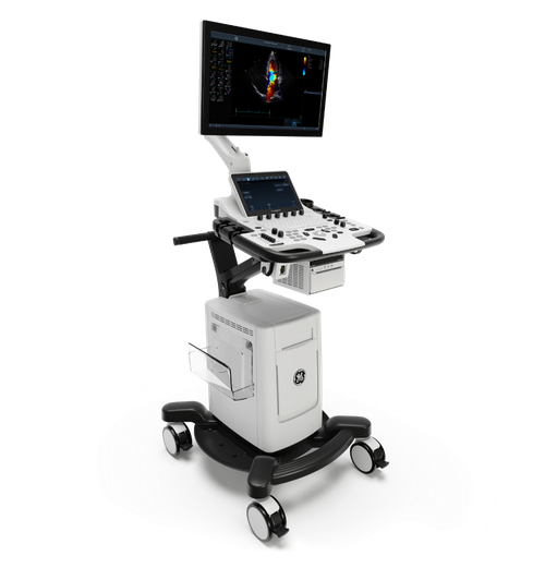
The GE HealthCare Vivid™ Explorer Cardiovascular Ultrasound System is a high-performance cardiac ultrasound platform designed for high-volume echocardiography, advanced cardiac assessment, structural heart imaging, and routine interventional procedures. Built on the same core technology foundation as GE HealthCare's ultra-premium Vivid Pioneer, Vivid Explorer combines exceptional 2D, multi-plane, and 4D imaging with cSound™ architecture, advanced cardiac quantification, AI-powered automation, and a comprehensive range of TTE, TEE, and ICE imaging options.
Designed to move seamlessly from the echo lab to interventional and electrophysiology environments, Vivid Explorer provides the imaging versatility and workflow tools needed for both everyday cardiovascular studies and more complex cardiac procedures.
Vivid Explorer combines 2D, multi-plane, and 4D imaging within a single cardiovascular ultrasound platform.
This allows the system to support routine transthoracic echocardiography while expanding into advanced TEE, structural heart, intracardiac, and interventional imaging workflows.
Its broad cardiac imaging capabilities make Vivid Explorer particularly well suited for facilities that need one system to serve both a high-volume diagnostic echo lab and procedure-oriented cardiovascular environments.
At the center of Vivid Explorer is GE HealthCare's cSound™ software-based imaging architecture.
cSound uses advanced processing to optimize contrast, spatial resolution, and tissue visualization, supporting consistent cardiac imaging across a broad range of patients and examination types.
Vivid Explorer shares its core technology foundation with GE HealthCare's Vivid Pioneer platform, bringing advanced Vivid imaging technology to a system designed for high-volume echocardiography and routine interventional use.
Vivid Explorer supports comprehensive 2D transthoracic echocardiography (TTE) for routine and advanced cardiac assessment.
The system can be used to evaluate:
Advanced processing and dedicated cardiac transducers support detailed visualization across a wide range of patients.
Vivid Explorer extends beyond conventional 2D echocardiography with comprehensive 4D cardiac imaging.
Volumetric imaging provides additional visualization of cardiac structures and can be particularly valuable when assessing complex anatomy, valves, ventricular function, and structural heart disease.
The system supports 4D imaging across multiple cardiac approaches, including compatible TTE, TEE, and ICE technologies.
Vivid Explorer supports multi-plane imaging, allowing clinicians to visualize cardiac anatomy from multiple imaging planes.
This capability is particularly valuable during transesophageal and interventional examinations where understanding the spatial relationship between cardiac structures can be critical for assessment and procedural guidance.
One of Vivid Explorer's defining capabilities is its broad transducer and imaging support across the cardiac care pathway.
The platform supports:
This allows Vivid Explorer to move beyond the traditional echo lab into structural heart and electrophysiology workflows.
The 4Vc-D volume transducer supports advanced transthoracic cardiac imaging on Vivid Explorer.
GE demonstrates the 4Vc-D for applications including:
This allows clinicians to transition between conventional and volumetric cardiac imaging using a compatible transthoracic probe.
The 6Sc-D is designed to support high-resolution 2D cardiac imaging across a broad patient population.
Its deep-penetration capabilities work with cSound processing to support detailed visualization in routine and challenging studies.
A slim, elongated probe design helps provide access through tight intercostal spaces, making it useful for smaller patients and challenging anatomies.
GE also demonstrates the 6Sc-D for pediatric cardiac imaging.
Vivid Explorer supports advanced transesophageal echocardiography using compatible TEE transducers.
GE demonstrates the system with probes including the 6VT-D and 9VT-D, supporting advanced visualization of cardiac structures and valves.
Compatible configurations can provide 4D TEE, allowing volumetric visualization during complex diagnostic and interventional cardiac examinations.
Vivid Explorer extends cardiac imaging into the catheterization and electrophysiology environment with intracardiac echocardiography (ICE) support.
The platform supports compatible 2D and 4D ICE technologies, allowing clinicians to visualize cardiac anatomy from within the heart during applicable catheter-based procedures.
Supported catheter technologies and functionality depend on system configuration and regional availability.
Vivid Explorer supports 4D intracardiac echocardiography with compatible ICE technology.
4D ICE provides volumetric intracardiac visualization that can help clinicians understand cardiac anatomy and device relationships during complex interventional procedures.
GE identifies compatible technologies including the 4D ICE NUVISION™ Ultrasound Catheter, subject to availability and regulatory status.
Vivid Explorer includes advanced capabilities designed specifically for structural heart assessment and procedural guidance.
These tools help clinicians visualize and quantify cardiac valves and other structures during pre-procedural planning and image-guided interventions.
Applications can include evaluation and planning involving the:
4D Auto MVQ (Mitral Valve Quantification) provides advanced analysis of mitral valve anatomy using 4D datasets.
The application is designed to streamline mitral valve visualization and quantification and can support assessment during planning for transcatheter mitral valve procedures.
GE specifically positions 4D Auto MVQ as a tool that can help assess TMVR screening.
4D Auto TVQ (Tricuspid Valve Quantification) provides automated visualization and quantification of tricuspid valve anatomy.
By applying automated analysis to 4D volume data, the application helps reduce the time and manual effort involved in evaluating complex tricuspid anatomy.
Vivid Explorer provides 4D Navigation capabilities for structural heart imaging.
The technology is designed to help clinicians reach desired 4D views more efficiently during complex cardiac assessment and procedural guidance.
This can help simplify navigation through volumetric datasets while reducing the manual effort required to establish clinically useful viewing planes.
Vivid Explorer supports tools for left atrial appendage (LAA) measurement and procedural planning.
These capabilities can assist clinicians in evaluating LAA anatomy and preparing for applicable structural heart interventions.
Availability of individual tools depends on system configuration and installed software.
AI Auto Measure 2D uses artificial intelligence to automate commonly performed 2D cardiac measurements.
By reducing the amount of manual interaction required for routine measurements, the technology can help streamline examination workflows and improve consistency between users.
Easy AutoEF automates the calculation of left ventricular ejection fraction.
The application is designed to simplify a commonly performed cardiac measurement, reducing manual tracing and allowing clinicians to obtain EF results with fewer workflow steps.
Easy AFI LV provides automated left ventricular strain analysis.
AI-based view recognition helps identify appropriate apical views and assists with myocardial analysis, supporting assessment of global and segmental left ventricular function.
AI Cardiac Auto Doppler applies automation to cardiac Doppler analysis.
The technology is designed to reduce manual tracing and measurement steps during routine Doppler assessment, helping standardize workflows and improve efficiency across examinations.
Vivid Explorer incorporates AI and automation throughout the cardiac imaging workflow.
These technologies are designed to help:
The goal is to allow clinicians to spend less time manipulating controls and measurements and more time evaluating clinical information.
GE HealthCare specifically positions Vivid Explorer for high-volume echocardiography environments.
The combination of high-performance imaging, AI-assisted measurements, automated quantification, customizable controls, and a familiar Vivid interface is designed to support efficient examinations throughout a busy clinical schedule.
Vivid Explorer is also designed to move beyond the echo lab into interventional and electrophysiology environments.
Its combination of TEE, 4D imaging, ICE, structural heart quantification, and navigation tools allows the platform to support applicable image-guided cardiovascular procedures.
This versatility can allow healthcare organizations to use a common Vivid platform across multiple stages of cardiac care.
Vivid Explorer incorporates the familiar Vivid workflow with an interface designed for efficient daily operation.
Features include:
These features help users personalize the system around their preferred scanning workflow.
An optional battery supports continued operation when Vivid Explorer is moved between clinical locations.
Battery-supported workflow can help maintain continuity when moving the system from:
Battery functionality is configuration-dependent.
GE HealthCare's current Vivid family is designed around a common technology foundation while providing different levels of cardiovascular imaging capability.
| System | Primary Positioning |
|---|---|
| Vivid Pioneer | Ultra-premium platform for the most complex cardiovascular imaging and interventional cases |
| Vivid Explorer | High-volume echo labs and routine interventional procedures with advanced 2D, multi-plane and 4D imaging |
| Vivid Advanced | Comprehensive cardiac imaging for busy hospitals and cardiology practices, plus vascular, abdominal and soft-tissue applications |
| Vivid Focus | Everyday cardiac imaging with streamlined 2D capabilities and broader general-imaging support |
Within this family, Vivid Explorer is the step below Vivid Pioneer for advanced cardiovascular imaging and provides significantly more advanced 4D and interventional capabilities than Vivid Advanced or Vivid Focus.
| Specification | GE Vivid Explorer |
|---|---|
| System Type | Cart-based cardiovascular ultrasound |
| Primary Specialty | Cardiology / Echocardiography |
| Imaging Architecture | cSound™ |
| 2D Cardiac Imaging | Yes |
| Multi-Plane Imaging | Yes |
| 4D Cardiac Imaging | Yes |
| TTE | Yes |
| 4D TTE | Yes |
| TEE | Yes |
| 4D TEE | Yes, with compatible transducer |
| Mini-TEE | Supported |
| ICE | Yes |
| 4D ICE | Supported with compatible technology |
| AI Auto Measure 2D | Available |
| Easy AutoEF | Available |
| Easy AFI LV | Available |
| AI Cardiac Auto Doppler | Available |
| 4D Auto MVQ | Available |
| 4D Auto TVQ | Available |
| 4D Navigation | Available |
| Structural Heart Imaging | Yes |
| Battery | Optional |
| User Interface | Customizable with tiltable touch panel |
Features, applications, transducers, and software capabilities vary by system configuration and purchased options.
Vivid Explorer supports a comprehensive cardiovascular probe portfolio that includes:
GE currently demonstrates Vivid Explorer with transducers including:
Exact transducer compatibility should be confirmed according to system configuration, software version, clinical application, and regulatory market.
The GE HealthCare Vivid Explorer is well suited for:
Get in the know to get the most value of your solution.